Ultrasonic Diagnostic Equipment: A Practical Step-by-Step Guide in 2026
What is it used for in 2026
Ultrasonic diagnostic equipment in 2026 plays a crucial role in various laboratory settings, primarily in medical diagnostics, veterinary practices, and research environments. The equipment is utilized for imaging soft tissues in real-time, aiding in the assessment of conditions such as cardiac abnormalities, gestational development in obstetrics, and organ evaluations in small animals.
History and evolution of the technology
The evolution of ultrasonic diagnostic equipment dates back to the early 20th century when sonar technology was primarily used in naval applications. Over the decades, as medical imaging grew in importance, ultrasound technology began to be adapted for clinical use. The development of portable and user-friendly devices, particularly in the last decade, has made it more accessible in various laboratory and clinical settings.
How to use it step by step
Using ultrasonic diagnostic equipment involves several key steps:
- Preparation: Ensure all necessary supplies and the device are ready for use.
- Patient/Subject Positioning: Position the subject appropriately for optimal imaging.
- Ultrasound Probe Selection: Choose the appropriate probe based on the specific application (e.g., linear or convex).
- Device Settings: Adjust the machine settings, such as gain and depth, according to the procedure.
- Image Acquisition: Apply a coupling gel to the area and move the probe to obtain images.
- Data Storage: Save the images and any necessary measurements for future reference.
- Report Generation: Generate and review reports based on the collected data.
Best techniques and protocols
For effective usage of ultrasonic diagnostic equipment, following these best practices is recommended:
- Utilize appropriate coupling gels to ensure optimal sound wave transmission.
- Maintain a steady hand while operating the probe for clear imaging.
- Regularly calibrate the equipment to ensure accuracy in diagnostics.
- Continuously update your skills and knowledge regarding new techniques and protocols in ultrasound technology.
Practical applications by laboratory type
Different laboratories apply ultrasonic diagnostic equipment in various ways:
- Veterinary Laboratories: Use it for cardiac assessments, reproductive health, and soft tissue evaluations.
- Medical Clinics: Employ ultrasonic equipment for obstetric imaging, organ assessments, and vascular studies.
- Research Facilities: Utilize it for anatomical studies and advanced imaging research.
Regulations, standards and certifications
Ultrasonic diagnostic equipment must comply with strict regulations and standards to ensure safety and efficacy. Regulatory bodies such as the FDA in the United States and similar organizations in other regions set forth guidelines that manufacturers must follow to certify their devices for clinical and laboratory use. Additionally, practitioners must be licensed and trained to operate these devices to meet legal and ethical standards.
Comparison with alternative technologies
While ultrasound is a widely used diagnostic tool, other imaging technologies such as MRI and CT scans offer different benefits and drawbacks. Ultrasound is particularly advantageous for its portability, cost-effectiveness, and real-time imaging capabilities, while MRI provides superior detail for soft tissue structures. Understanding these comparisons helps practitioners choose the best equipment for specific diagnostic needs.
Comparison of available models
| Model | Best for | Key specs | Recommended use case |
|---|---|---|---|
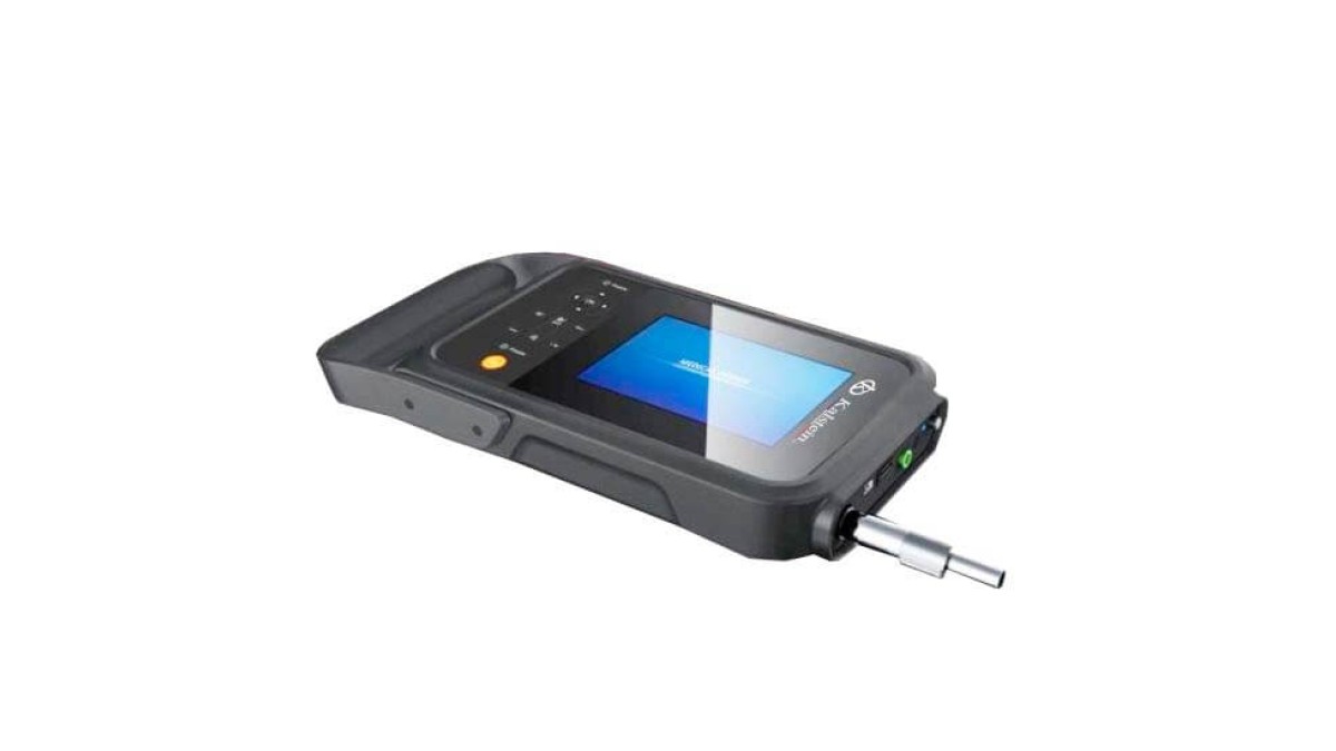
| YR05146 | Veterinary diagnostics | Weight: 0.5 kg, 7.0" LCD, 1024 images | Portable use in veterinary practices |
| YR05147 | Obstetric and gynecological examinations | 15" LED monitor, 6 automatic report types | GYN and obstetrics procedures |
| YR05154 | General diagnostics | Price: $850.00 | Routine ultrasound examinations |
| YR05154-1 | Tablet integration | Price: $1150.00 | Mobile diagnostic solutions |
| YR05155 | Linear imaging | Price: $950.00 | Musculoskeletal and vascular assessments |
| YR05155-1 | Advanced tablet use | Price: $1190.00 | Field diagnostics and assessments |
Common mistakes and how to avoid them
Practitioners can often make several mistakes when using ultrasonic diagnostic equipment:
- Not using adequate gel can lead to poor image quality—always ensure proper application.
- Neglecting to calibrate the machine regularly may lead to inaccurate results.
- Improper patient positioning can obstruct clear imaging—always verify positioning before starting.
Maintenance, calibration and good practices 2026
To ensure the longevity and performance of ultrasonic diagnostic equipment:
- Regularly clean probes and components according to the manufacturer's instructions.
- Schedule routine maintenance checks to assess the functionality of the device.
- Keep software updated to leverage the latest features and improvements.
Cost-benefit analysis 2026
When considering ultrasonic diagnostic equipment, performing a cost-benefit analysis is essential:
- Evaluate initial costs against long-term savings on diagnostic procedures.
- Assess the potential increase in patient throughput due to faster imaging capabilities.
- Consider the range of applications possible with the device, which can justify higher upfront costs.
Frequently asked questions
What should I consider when choosing ultrasonic diagnostic equipment?
When choosing ultrasonic diagnostic equipment, consider factors such as the specific applications required, portability, ease of use, and compatibility with existing systems in your laboratory.
How often should I calibrate my ultrasound machine?
It is recommended that you calibrate your ultrasound machine at least once a year or more frequently if you notice any discrepancies in imaging quality.
What are the best practices for patient preparation before an ultrasound?
Ensure that the patient understands the procedure, is comfortably positioned, and has followed any necessary pre-examination instructions, such as fasting if required.
How can I improve the imaging quality of my ultrasound equipment?
Improving imaging quality can be achieved by ensuring proper gel application, adjusting machine settings, and maintaining optimal patient positioning during the scan.
What types of reports can I generate from ultrasonic diagnostic equipment?
Most modern ultrasound machines offer various automated reporting options, including measurements and assessments for a range of specialties such as obstetrics and cardiology.
Can I integrate ultrasound machines with other diagnostic systems?
Yes, many ultrasound machines are designed with connectivity options that allow integration with additional diagnostic systems, enhancing data management and workflow efficiency.
How can I request a quote for ultrasonic diagnostic equipment?
You can easily request a quote by contacting our sales team or visiting our website to explore our catalog of ultrasonic diagnostic equipment and receive personalized assistance.
If you are looking for a fusion of innovation and quality, you have come to the right place. At Kalstein, we offer you the luxury of exploring our exclusive catalog of laboratory equipment. We manufacture every device to the highest standards of excellence. Our intuitive and seamless online purchasing channels are designed for your convenience, securing the most competitive prices. Hesitate no longer — we bring science to life, it is time to become part of our community.