Color Doppler Ultrasound Cart with Wheels 3D Model YR05149
Color Doppler Ultrasound Cart with Wheels 3D Model YR05149 — Kalstein laboratory equipment with technical specifications, advanced features, and certified professional solutions for scientific use.
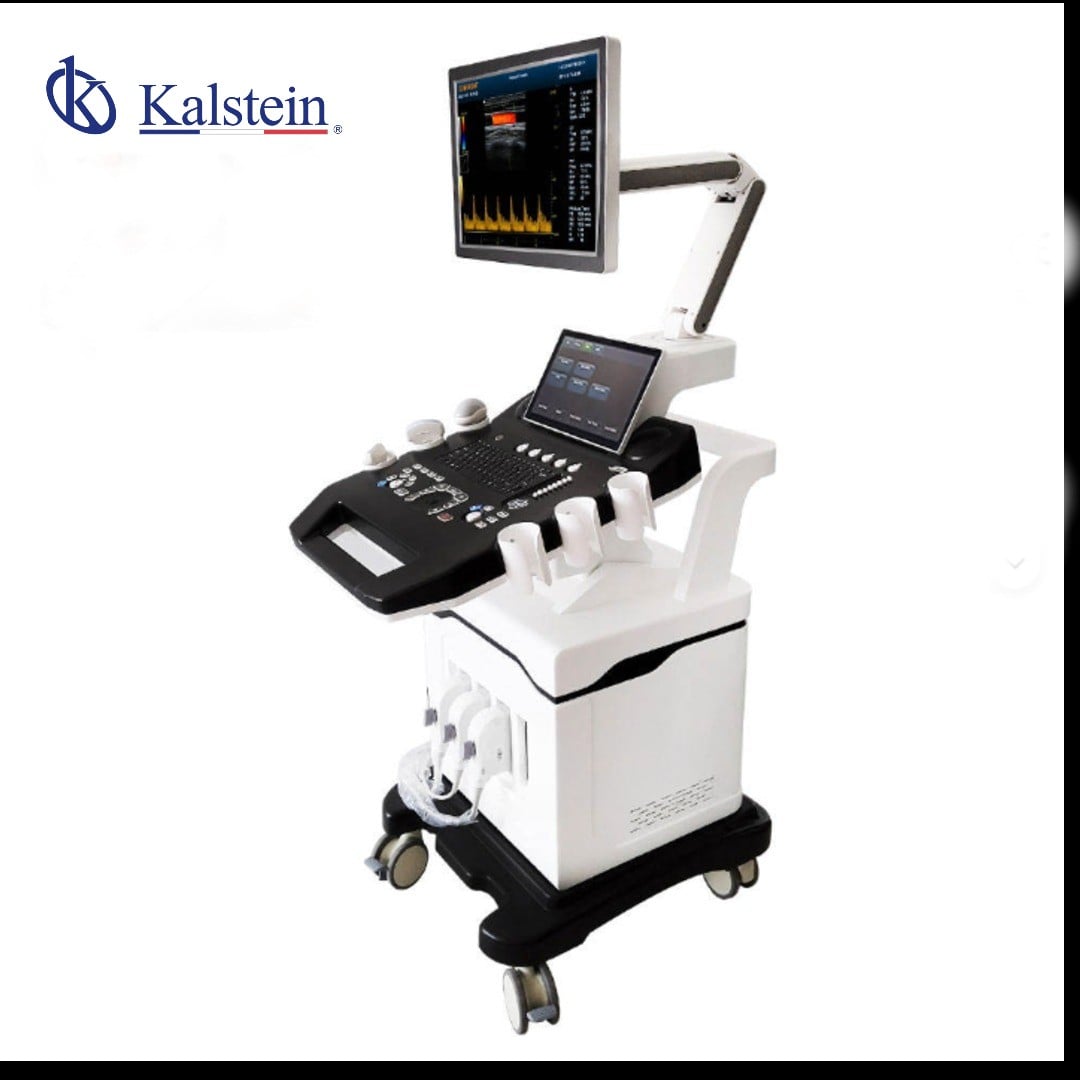
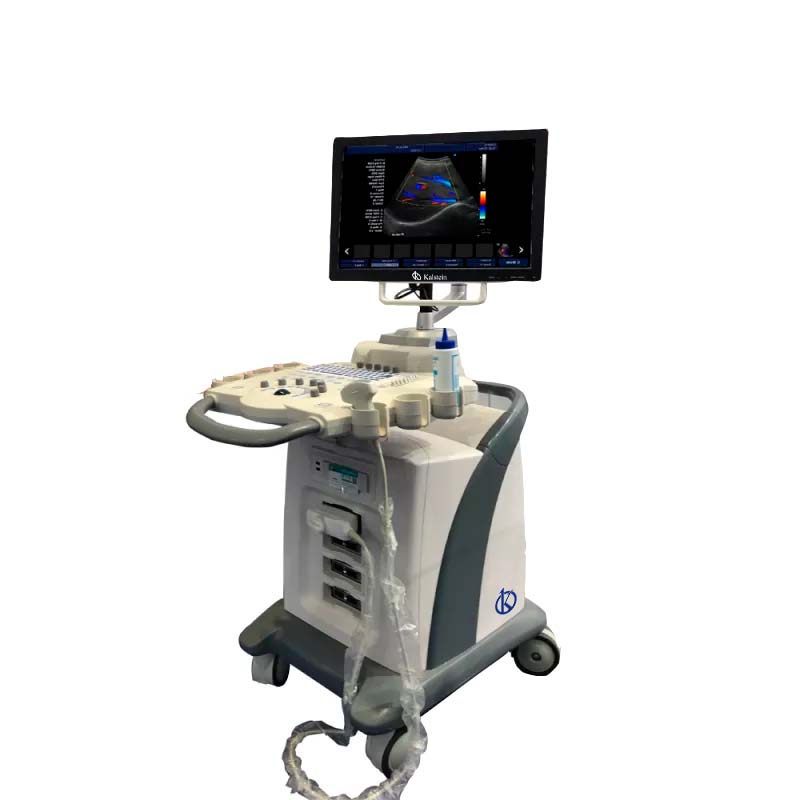
The Color Doppler Ultrasound Cart with Wheels 3D Model YR05149 offers cutting-edge imaging technology packed into a highly mobile and versatile unit suitable for various medical environments. Equipped with a 15-inch LED screen featuring multilingual support, this ultrasound cart ensures clear, precise imaging for accurate diagnostics. The model supports multiple imaging modes such as CF+B, PDI, DPDI, and others, allowing for comprehensive anatomical and physiological assessments. Additionally, it boasts user-friendly features such as automatic measurements, integrated DICOM 3.0 protocol, and user rights control for secure use. With its built-in online instructional feature, this model guides users step-by-step through operation, making it ideal for both expert professionals and healthcare facilities aiming for enhanced productivity.
Market Price
The price for ultrasound machines such as the Color Doppler Ultrasound Cart with Wheels can vary widely depending on features and brand reputation. On the market, these machines typically range from $20,000 to $50,000. The YR05149 model, with its advanced features and robust construction, is positioned competitively within this range. For specific pricing and potential discounts, you can request a quote on the Kalstein Plus platform.
Frequently Asked Questions
What types of imaging does this ultrasound support? The machine supports a variety of imaging modes including trapezoidal imaging, virtual convex matrix, and harmonic imaging, among others.
Can I connect it to external devices? Yes, the system provides multiple connectivity options such as VGA, HDMI, and USB ports for easy integration with peripherals.
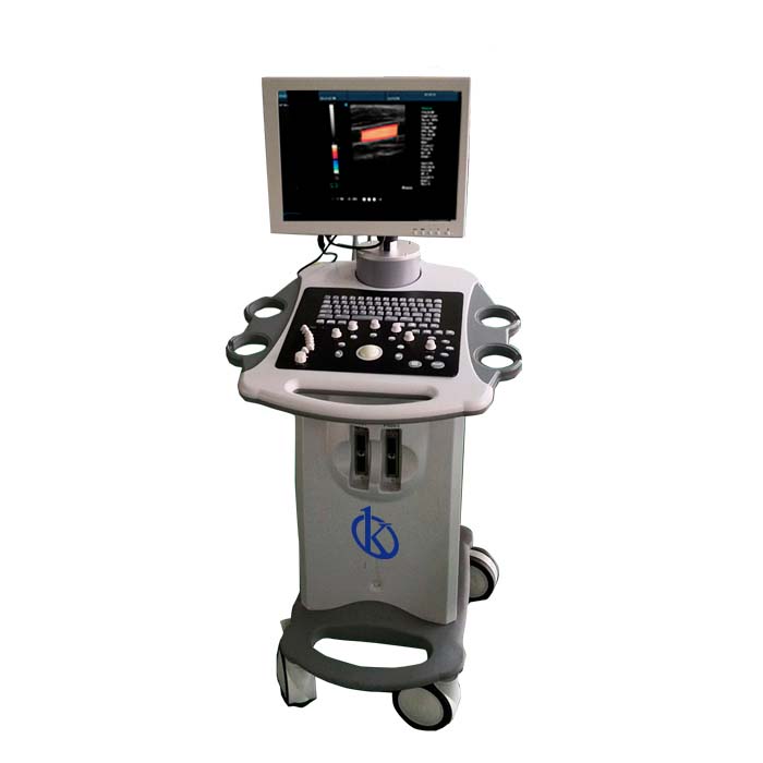
Is it easy to move the cart? Yes, with its builtin wheels, it's designed for easy mobility across different medical rooms and settings.
Advantages and Disadvantages
Advantages: The YR05149 model is multifunctional, providing high-quality imaging across several modes. Its user-friendly design, coupled with the automatic features, enhances diagnostic efficiency. The multilingual support also broadens its usability in diverse international settings.
Disadvantages: As a high-tech device, it requires initial setup and understanding of its advanced functions, which might pose a learning curve for new users.
Product Usage in the Field
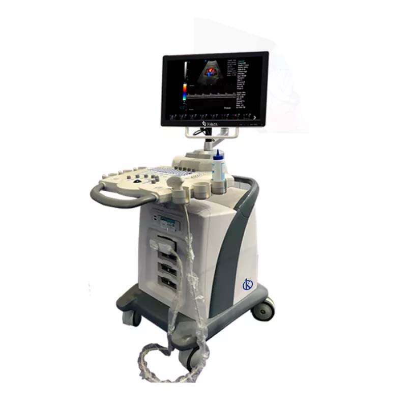
This model is particularly effective in hospital radiology departments, clinics, and private practices for cardiovascular assessments, obstetric imaging, and general ultrasound diagnostics. Its capable of delivering clear images even in complex cases, enhancing the accuracy of clinical evaluations.
Recommendations
To maximize the utility of the Color Doppler Ultrasound Cart YR05149, it is recommended to regularly update the software to ensure access to the latest features and security enhancements. Proper training for the medical staff will also optimize the functionality of this advanced imaging device.
Features
- 15' LED screen with multilingual support
- Advanced imaging modes including CF+B, PDI, DPDI
- Automatic measurements and user rights control
- Integrated DICOM 3.0 protocol
- Built-in online user guidance
Technical Specifications
| Model | YR05149 | |||
| Connectivity/Media/Peripherals | ||||
| Transducer Ports | 4 | |||
| USB ports | 4 | |||
| HDD | 64GB (SSD), 120G/200GB SSD (optional) | |||
| Foot switch | USB | |||
| Ethernet port | 2(100Mb/1000Mb) | |||
| External screen | VGA,HDMI, | |||
| Printer (Optional) | USB Printer, Digital Laser Printer, B/W Digital Thermal Printer | |||
| Printing area | Image, report, Image+report | |||
| Cine/Picture Memory | ||||
| Memory Cinema | 1200 frames (max) | |||
| Film Review Speed | 1, 2, 4, 8 | |||
| Cinema Review Loop | YES | |||
| Capture function | YES | |||
| DICOM connectivity | DICOM3.0 Compliant | |||
| 3D software | Built-in 3D software | |||
| Image Storage | Storage Format: PNG, AVI, BMP, JPEG, DICOM Export Video Format: AVI Export Image Format: PNG, JPEG, BMP, DICOM USB Flash Drive | |||
| Technology | ||||
| Panoramic Imaging Technology Full Digital Signal Processing Technology Multi-Beamforming Technology Spot Reduction Technology Tissue Harmonic Imaging Dynamic Tissue Optimization Technology Duplex and Triplex Synchronous Display Directional Power Doppler Imaging Parameters Imaging Parameters special tissue presets PW Auto Trace Update online CF+B mode on one screen Complex model imaging Automatic IMT measurements Virtual Convex Array Trapezoidal imaging | ||||
| Overall Performance | ||||
| Digital Broadband | 12288 channels | |||
| Beam Former | reprogrammable | |||
| Transmission voltage | Adjustable (15 steps) | |||
| Beamformer Frequency Range | 1~40MHz | |||
| LED monitor | ||||
| Size (diagonal) | fifteen" | |||
| Contrast Ratio | 800: 01: 00 | |||
| Resolution | 1024×768 pixels | |||
| Brightness | 230 cd/m2 | |||
| Color Depth | 24 bit | |||
| Rotation Angle | ±90° | |||
| Gray levels | 256 | |||
| Imaging performance | ||||
| Start Time (max.) | Average < 90 seconds | |||
| Preset Switching Time | Average < 1 second | |||
| Storage Time (Image to disk) | Average < 0.5 seconds | |||
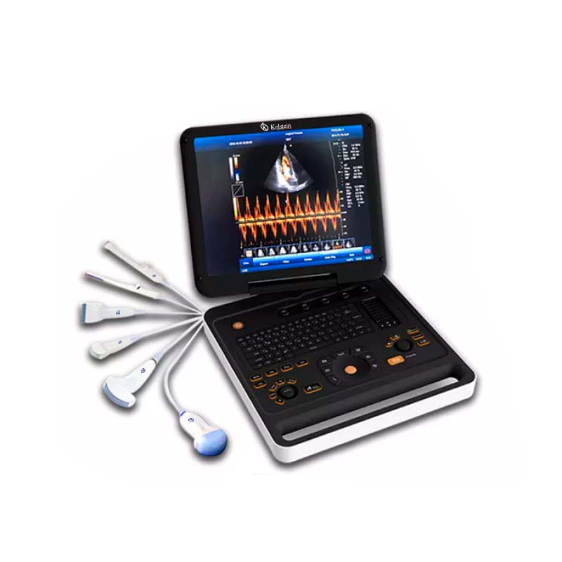
| Transducers | ||||
| Research | Convex array probe | Linear Array Probe | Intracavitary probe | Microconvex probe |
| Frequency | Center 3.5MHz | Central 7.5MHz | Center 6.5MHz | Center 4.0MHz |
| (2.0MHz to 10.0MHz) | (6.0MHz to 10.0MHz) | (5.0MHz to 9.0MHz) | (2.0MHz to 5.5MHz) | |
| Field of play | 0.516mm | 0.352mm | 0.216mm | |
| Radio | 60mm | N/A | 10mm | |
Technical specifications
| Dimensions | L 162 × W 72 × H 95 cm |
|---|---|
| Weight | 222 kg |
| Manufacturer | Kalstein |
Video Color Doppler Ultrasound Cart with Wheels 3D Model YR05149
Frequently asked questions
How to know the prices of Color Doppler Ultrasound Cart with Wheels 3D Model YR05149?
To know the price of Color Doppler Ultrasound Cart with Wheels 3D Model YR05149, please send us an email with your request through the contact form AQUI.
What are the delivery times for Color Doppler Ultrasound Cart with Wheels 3D Model YR05149?
Delivery time depends on stock availability and freight type (air or sea). In stock: air 15-30 days, sea 45-60 days. Out of stock: air 30-60 days, sea 60-90 days.
How to make a purchase of Color Doppler Ultrasound Cart with Wheels 3D Model YR05149?
You can buy by email ([email protected]), phone (+33 (0) 1 70 39 26 50) or through the official Kalstein website in your country.
How does the warranty work for Color Doppler Ultrasound Cart with Wheels 3D Model YR05149?
All Kalstein equipment comes with a 1-year warranty against manufacturing defects. The warranty does not cover damage from improper installation or misuse. See our «terms and conditions» AQUI.
Can I request a quote online for Color Doppler Ultrasound Cart with Wheels 3D Model YR05149?
Yes, you can request a quote for the Kalstein equipment you are interested in directly from our official website. Click AQUI.